Retinal Artery Occlusion
Greetings to all and sundry,
And happy new month to us all, it is my fervent hope that March blesses us with loads of goodwill and also that the crypto market would soar higher and higher. As usual new week marks new stress and workload and the new month comes with its own goodness and challenges, irrespective let's continue to give our best in everything we do.

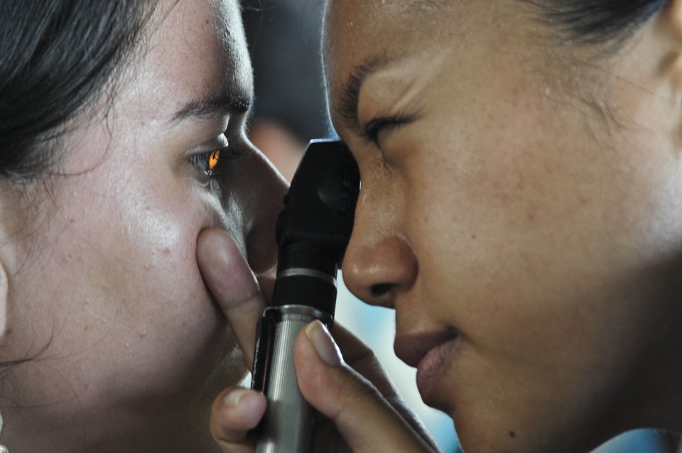
So far the week has been good at my end and I am very much optimistic that it has been great for my readers as well, I do hope you are all well and ready for the new month. So today I am here at your doorstep and I am sure that now when you see my write-up the first thing that comes to mind is the eye? Well, you are right, as an Optometrist, my passion is about ocular health, well music too is part but for today, let's have another lesson about our eyes.
Introduction
The eye is not so much of a big organ as compared to others within the body, and yet, it is quite powerful and has such an important function that when it is affected or not functioning one's entire life could literally come to halt. Because of the small nature of the eye in terms of space it occupies and its size, most of the anatomy and physiological features that interact with it tend to be small even if naturally they are macro.
Without a size reduction, they may not be able to execute whichever function or duty when it comes to the eye. One such feature is the blood supply that goes to the eye. Much like any other part of the body, the eye also receives innervations from the nervous system and vasculature from the cardiovascular system. The innervations ensure that vision is made possible, and pain and other equally important features related to the nervous system are felt.

Regarding vasculature, the eye has to receive nutrition and oxygen from the body to be able to survive and continue functioning accordingly. As a matter of fact, the cells in the eye, more specifically the retina (the part of the eye responsible for vision using changing light energy to electrical impulses) have one of the highest metabolic rates such that when they are deprived of oxygen or ATP for just some few hours they tend to die off permanently which results in permanent loss of vision concerning the affected cells.
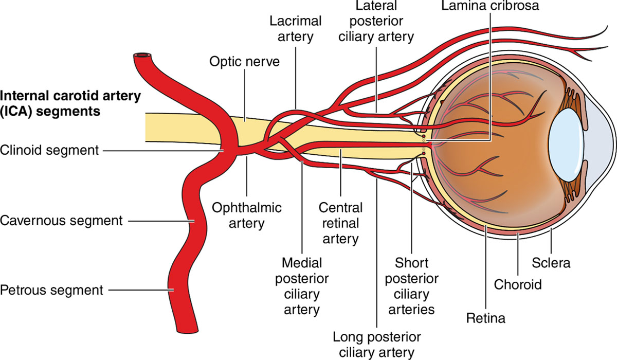
This goes on to emphasize how important vascularization is important when it comes to the eye. And so when oxygenated blood is pumped from the heart, some of it enters the ophthalmic artery after the internal carotid artery, which supplies areas within the head region, including the brain. The ophthalmic artery then sends some to the central retinal artery which is basically the main supplier to the eye which then feeds the retina. And so when something happens to this artery, all hell breaks lose, for our discussion today though, this artery is going to be our focal point.
Retinal Artery Occlusion
Now when the central retinal artery enters the eye through the optic disc area called lamina cribrosa (do not focus on the technicalities, focus on the understanding of the general condition) it splits up so that it can feed the various branches. Remember how we talked about vessels having to be micro because the eye is small and you need to come down to its level to interact with it? This is where it plays out.
The more these vessels branch out, the small or more micro they become. This makes it quite easier for embolus or other factors such as glucose to get lodged within the vessels, cutting off blood passage through it and thus blood supply to specific areas of the retina. When this happens we say that the artery has been occluded. Now let me quickly remind you that we also mentioned previously that the cells of the retina are so active that when they don't get oxygen which a short time they die permanently. And so putting this together, I hope you are getting how serious retinal artery occlusion is now?

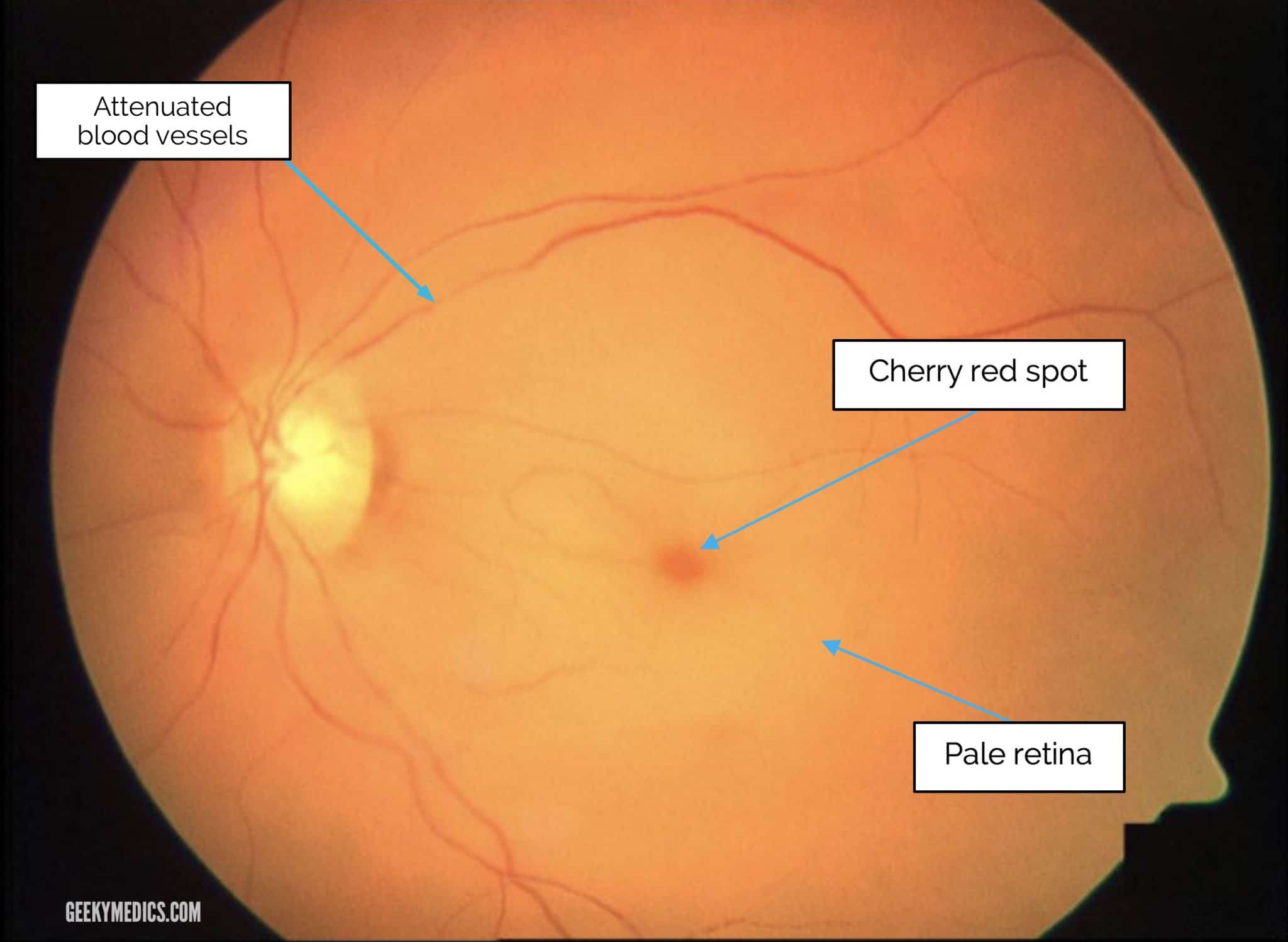
It is more terrible if the occlusion occurs at the level of the central retinal artery, why? Because this is the main supplier that would branch off the supply the others and so basically the entire retina would have to die off within a very short while if supply is not re-established within a matter of a few hours. And so sudden painless loss of vision could easily be attributed to central retinal artery occlusion. The lack of oxygenated blood and hypoxia would cause edema on the retina as well as a white appearance (this would be seen by your optometrist) signifying the death of cells.
If you are lucky enough to have the occlusion occurring within a branch of the central retinal artery then the same thing would happen however it would be localized to a particular area of the retina which means, loss of visual fields, which a better option to loss of the entire peripheral retina though. Now you may notice how I suddenly added peripheral to the retina? Let's explain that. A part of the retina called the macular which is the part used for central vision tends to get its supply from vessels within a layer behind it called the choroid. These vessels are called chorioretinal arteries and feed the macular from behind.
Because of this, in the event of either a central retinal or branched retinal artery occlusion, the macular gets spared because of the alternate source of nourishment, this thus ends up producing a cherry red spot appears in your eye where all other parts of the retina look very pales with the middle or area of the macular look reddish and standing out amongst them. Retinal artery occlusion is actually considered an emergency and it is a condition that must be sought to resolve within the shortest possible time lest the consequences are dire.

In trying to make things better for the eye, the focus of care is on trying to dislodge whatever may be blocking the vessel, whether it be embolus or glucose (this is seen in diabetic retinopathy situations). Over the years studies and research have shown that trying to bring down the pressure within the eye can help, this may be done with the use of an IV injection of mannitol or massaging the eye.
Vasodilators and anticoagulants also tend to help, breathing into a polythene bag in this instance may also be helpful. Hopefully, any of these can get the blood flow restored and thus save the vision of the patient, if not then permanent damage may be expected. Do not get alarmed if you notice a sudden painless loss of vision though, there are a few other things that could also cause this and over time more of these would be explored, for now, the advice is to consult your primary eye care physician right away if you notice anything out of place with regards to your eyes or vision.
Conclusion
In this instance, how quickly you go in for consult may just make all the difference in you being able to save your sight or losing a chunk of your vision permanently. It is my fervent hope that none of us get to experience this, however, please do not hesitate in seeing your Optometrist if you notice anything with your vision especially when you fall in the risk factors group of diabetes, hypertension, obesity, drinking, and the likes.

You only have one life and one sight or two sights if you are counting the two eyes separately and whiles it is good news that his condition is mostly unilateral, it can be bilateral in some instances however, it wouldn't be advisable to play Russian roulette with your sight. And so I would wrap up here and say thank you for this opportunity and for taking the time to read what I had to share today. Please feel free to share your questions or comments with me, I appreciate your time and feedback, thanks once again and I wish you a marvelous day and month. Cheers!
Further Reading
Feltgen, N., & Pielen, A. (2017). Retinaler Arterienverschluss [Retinal artery occlusion]. Der Ophthalmologe : Zeitschrift der Deutschen Ophthalmologischen Gesellschaft, 114(2), 177–190. https://doi.org/10.1007/s00347-016-0432-4.
Hayreh S. S. (2018). Central retinal artery occlusion. Indian journal of ophthalmology, 66(12), 1684–1694. https://doi.org/10.4103/ijo.IJO_1446_18.
Scarinci, F., Cacciamani, A., Ripandelli, G., & Parravano, M. (2022). Branch retinal artery occlusion caught in the act by an optical coherence tomography angiography image: a case report. BMC ophthalmology, 22(1), 303. https://doi.org/10.1186/s12886-022-02517-5.
Dattilo, M., Biousse, V., & Newman, N. J. (2017). Update on the Management of Central Retinal Artery Occlusion. Neurologic clinics, 35(1), 83–100. https://doi.org/10.1016/j.ncl.2016.08.013.
Congratulations @nattybongo! You have completed the following achievement on the Hive blockchain And have been rewarded with New badge(s)
Your next target is to reach 2000 comments.
You can view your badges on your board and compare yourself to others in the Ranking
If you no longer want to receive notifications, reply to this comment with the word
STOPTo support your work, I also upvoted your post!
Check out our last posts:
Support the HiveBuzz project. Vote for our proposal!
Thanks for your contribution to the STEMsocial community. Feel free to join us on discord to get to know the rest of us!
Please consider delegating to the @stemsocial account (85% of the curation rewards are returned).
Thanks for including @stemsocial as a beneficiary, which gives you stronger support.