Structure of the Muscle || Sarcomere, Actin, and Myosin
I started writing on the muscles and I wrote on the function of those muscles. Today, we will be looking at the structure and function of different skeletal muscles. Muscle tissues are completely different from other tissues in the body, and they have a few characteristics that make them unique and they are; It ability to be stimulated, excited, or activated by motor neurons causing a change in membrane potential, With the effect of motor neurons, muscles can contract and also be stretched beyond its normal resting length in what is known as extensibility, and muscles are elastic, and always want to recoil. This said, if you were asked what the functions of muscles are, what would be your response? Muscles have certain functions which includes the ability to initiate locomotion/movement, Stability and posture against gravity, Stabilization of body joint, and generation of heat.

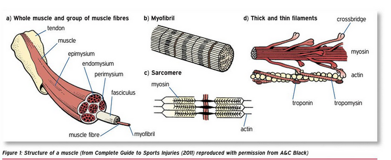
To explain the muscle structure, I will explain using a transverse cut. The muscle has a connecting tissue over its belly, known as the epimysium which is a dense fibrous irregular connective tissue which is connected to the muscle belly. The epimysium also attaches to the bone forming a site of attachment for some muscles. In the muscle belly are long muscle fibers known as fascicles which are sorrounded by a connective tissue known as perimysium and it is similar to the epimysium as it is dense, and ireegular. In the fascicles are muscle fibers/cells which makes it up, and each of these fibers have areola connective tissues known as endomysium. The endomysium is not dense or tough like other muscle fibers, and it covers the sarcolema.,.
A tendon is a connective tissue rich in collagen that connects the muscle to the bone.
When muscles contract, all muscle fibers contract as well. It pulls the endomysium, perimysium, epimysium, and the fascicles. When this connective tissues are pulled, they pull the tendon which then moves the bone in the direction of the contraction and the insertion of the muscles. When a muscle contract, the origin of the bone would not move while the insertion moves. When a mmuscle contracts, it moves the bone from insertion to origin, and this is done by the tendon which pulls the sheet (aponeurosis) that conects the muscle to the bone and they contribute to the elasticity of the muscles as they help it regain its structure. ,. The muscle to bone connection can be either through direct attachment or indirect attachment. Direct attachment of the muscle isn't common but the indirect attachment is very common as most of the ways the muscles connect to the bone is via indirect attachment. In the direct attachment, the epimysium attaches to the bone, along with the periosteum. When the periosteum, or perichondrium are attached to the bone and fuses to the epimysium, it is regarded as a direct attachment. Indirect attachment of muscles are mediated through tendons and aponeurosis. Tendons are small and can use up better space with indirect attachment, and prevent the destruction of the muscles when there is movement as tendons possesses collagens making it tough.,,.In the endomysium are myofibril which are made up of proteins, and in the muscle cells are sarcoplasmic reticulum which stores calcium in the muscles. The muscles cells are straited and it is made up of the Sacomere structure..

The Sarcomere is made up of proteins where from one z-disc to another z-disc is a functional unit of the muscle fiber, made up of protein molecules alpha actinin. In the sacomere, there are structures which include the thick filament which anchors to the z-disc by the titin, which runs through the thick filament and connect to the m-line which runs across the thick filament. The M-line is made up of Myomesin, C-proteins, and Creatine Kinase. From one end of a thick filament to another end, it is referred to as the A-Band (Anisotropic).,. Between the end of a Z-disc and another Z-disc is the Isotropic band (I-band) as it is a lighter band. The thick filament also possesses alpha-helix tails which are known as Myosin which is made up of the tail, the neck and the head. The head binds into the active site of the actin, allowing for the movement of the myofilaments, also the head can break down ATP into ADP in an inorganic phosphate, with the help of its enzyme component known as the Myosin ATPase. ,. At the top of the sacomere is the thin filament which is directly connected or anchored to the Z-disc through the nebulin. In the thin filament is another protein known as actin (G-actin when in monomer and F-actin when in filament as a result of polymerization..,. The myosin head is prevented from binding through the active sites, with the help of a protein called TropoMyosin when the myosin us at rest. Another protein molecule in the thin filament which binds to Actin, tropomyosin, and calcium is known troponin. The binding area with calcium is known as Troponin-c, the site where it binds with tropomyosin is known as Troponin T, and the site where it binds with actin is known as Troponin I..
Having explained the structure of the muscles, and taking a look at the sacomere structure which includes actin and myosin, talking about the functions of the proteins in the muscles will be easily understood and nueromolecular transmition in the skeletal muscle, as well as muscle mechanics.
Image 1 || Anatomy & Physiology by CCCOnline || Muscular Levels of Organization
Thanks for your contribution to the STEMsocial community. Feel free to join us on discord to get to know the rest of us!
Please consider delegating to the @stemsocial account (85% of the curation rewards are returned).
Thanks for including @stemsocial as a beneficiary, which gives you stronger support.
Congratulations @eni-ola! You have completed the following achievement on the Hive blockchain And have been rewarded with New badge(s)
Your next target is to reach 55000 upvotes.
You can view your badges on your board and compare yourself to others in the Ranking
If you no longer want to receive notifications, reply to this comment with the word
STOPCheck out our last posts:
Support the HiveBuzz project. Vote for our proposal!