Another Child Born With a Heart Defect || Acyanotic Congenital Heart Disease
Every baby is born beautiful, the gray or blue color of their eyeballs make them adorable, although, melanocytes still have a lot of work to do to make it get its color. Anyway, I am not here to talk about the coloring of the babies, instead I will be looking at why infants could have Congenital heart disease, and what could happen. When a child has a Congenital heart disease, it isn't the fault of the parents, the doctors, or even the baby. The child is born this way, since the abnormality occurred during fetal development.
As a parent, getting news about your baby having a heart defect which isn't your fault, and definitely not their fault, can be very disheartening. Every method which would help solve these defect would be considered because no parents want their children to die. When children have these heart defects, they could be Cyanotic (blue baby), Acyanotic. In this post, I will be explaining the Acyanotic Congenital heart disease in the very best way, and will focus on Cyanotic Congenital heart disease in our next post

Please sit back, grab a popcorn, and soda, and enjoy this reading...Oh, you should be learning not relaxing, so taking note is better
One is Colorful, the Other isn't
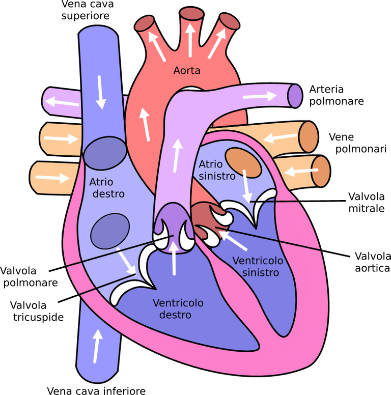
To explain the heart defect, let me do a small anatomy of the heart, and how blood flows in the heart. The heart, has four main chambers, which are divided into two sides vertically, and horizontally. When divided horizontally, we have the right and left Atrium, as well as the right and left Ventricle, and when divided vertically, it is the right ventricle and Atrium in one side, and the left Atrium and Ventricle on the other side. The Heart is made up of Atrioventricular valves, which are within the heart. Within the right Atrium and Ventricle is the Tricuspid valve, which prevents the blood being pushed from the right ventricle to go back into the right Atrium. Also, between the Left Atrium and the left Ventricle is the Bicuspid valve (Mitral valve) which prevents the back flow of blood into the left Atrium from the left Ventricle. Beneath the right ventricle, there is a valve that connects the right ventricle to the pulmonary trunk, known as the Pulmonary valve. The pulmonary valve (pulmonic valve) prevents blood from going back into the right ventricle. Furthermore, just between the left ventricle and the Aorta is the Aortic valve, which prevents the backflow of blow from the Aorta to the Left Ventricle during Diastole. In the exit and entry of blood into the heart, it should be clear that veins are bringing bloods into the heart, while arteries take blood away from the heart. The deoxygenated blood entering the heart first through the Superior (upper part)/Inferior (lower part) Vena Cava, gets into the right Atrium, and moves to the right Ventricle through the tricuspid valve. With the help of the pulmonary valve, the blood will exit the right ventricle into the lungs through the Pulmonary artery/trunk for oxygenation. From the lungs, oxygenated blood goes back to the heart through the Pulmonary Veins. It gets into the Left Atrium, then moves into the left ventricle via the Bicuspid valve. The Left ventricle pumps the blood to the aorta, which then sends it through the arteries into the body.
Congenital heart defects can be divided into two classes, which are the Cyanotic and Acyanotic defects. With Cyanotic defects/critical congenital heart disease, the heart pumps deoxygenated/less than usual oxygenated blood into the body. The heart experiences right-to-left shunt where the deoxygenated blood from the right heart, mixes with oxygenated blood from the left heart, which is then pumped into the body, leading to Cyanosis of the skin (a blue discoloration of the baby's skin), in babies, it is known as Blue baby. Acyanotic defect on the other hand doesn't affect the flow of blood, since it is always left-to-right shunts. With left-to-right shunts, oxygenated blood in the left heart mixes with deoxygenated blood in the right heart, thereby allowing the heart to send oxygenated blood to the lungs.
The No Cyanotic Conditions
The heart doesn't just become faulty, unless it has defects. In this case, the defects aren't going to cause the body to show bluish color as a result of deoxygenated blood being distributed around the body. In this case, the heart distributes oxygenated blood to the body. These defects include Atrial Septal defect (ASD), Ventricular Septal Defect (VSD), Patent ductus arteriosus (PDA),and Endocardial Cushion Defect (ECD).
With Atrial Septal defect (ASD), there is a hole between the inter atrial septum of the heart, leading to the shunting of blood from the left Atrium to the right Atrium. (Remember that the left Atrium and Ventricle has higher pressure compared to the right Atrium and Ventricle). The blood that goes into the right Atrium, from the left Atrium, is oxygenated, and this is why the body wouldn't experience Cyanosis. If it were in the case where deoxygenated blood was being pumped into the Aorta, then cyanosis would have happened.
The Ventricular Septal Defect (VSD), has to do with a hole/opening in the septum of the Ventricles in the heart. This opening allows blood to shunt from the left ventricle which has a higher pressure to the right ventricle which has a lower pressure. Since the defect is causing the movement of oxygenated blood, to the right ventricle, then no Cyanosis is experienced.
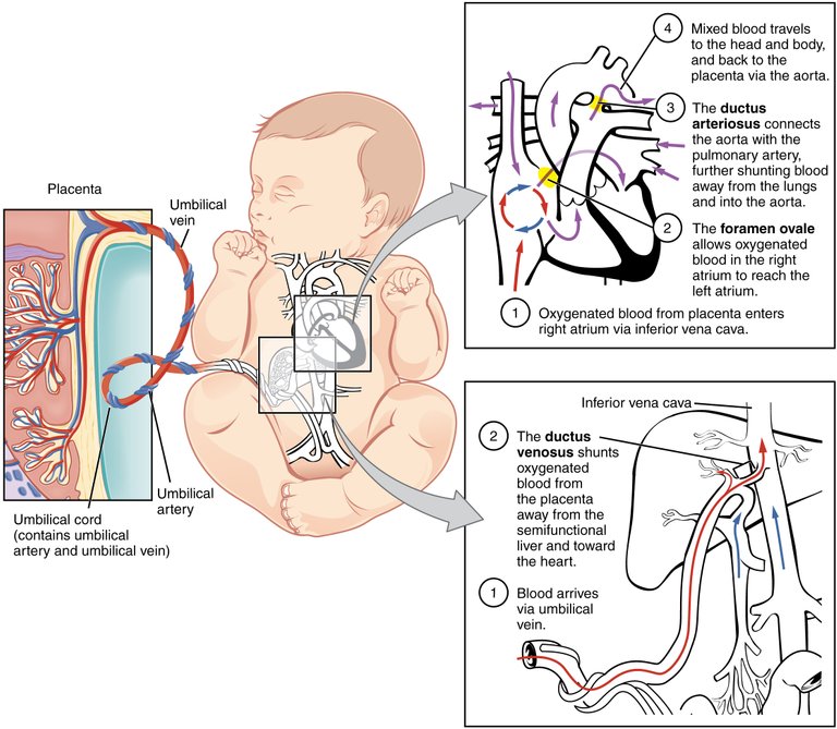
With patients suffering from Patent Ductus Arteriosus (PDA), the doctus arteriosus (in infants) which is a hole between the aorta and the pulmonary artery doesn't close after birth, thereby causing the shunting of oxygenated blood from the aorta to the pulmonary artery.
The next being the Endocardial Cushion Defect/Atrioventricular Septal Defect, is a defect with the heart where there is a hole in the septum of the Left Atrium, and right Atrium, thereby allowing blood to shunt from the Left Atrium to the right Atrium, and there is also a hole between the septum of the ventricles, thereby causing blood from the left ventricle to shunt into the right ventricle as well. In other word, the intravascular wall separating the four chambers of the heart is either poorly formed, have holes, or is absent. But with Acyanotic Congenital heart defects, the left part of the heart is the one shunting blood into the right side of the heart.
In a Normal Atrial wall in a fetus heart, the septum primum goes the entire lenght to the septum intermedium, which is the line between the septum of the Atrial and the Septum of the Ventricles. In the Septum Primum, there is a hole known as the Ostium Secundum which covered by the Septum Secundum which separate the atrails of the heart in the infant. Still in the septum dividing the atrial, is the Foramen Ovale, an opening which usually closes to become scar tissue, months after the child is born.. A defect in any of these could lead to a Congenital Heart Disease, including the Primus Atrial Septal defect, and Secundum Atrial Septal defect.
In a normal ventricular wall of an fetus heart,the interventricular septum which seperates the two ventricles, joins the septum intermidium. The Interventricular Septum has two parts, which are the muscular portion, and membranous portion. A defect in the parts of the interventricular septum can cause Congenital heart defects such as the Membranous Ventricular Septal Defect, Muscular VSD, Atrioventricular canal type VSD, and Conal septal VSD..

For Every Action, There is an Equal and Opposite Reaction
Understanding why children would have these Acyanotic Congenital Heart Disease will be important in this post, or don't you think so?
Infants with these disorders could be as a result of Chromosomal abnormalities. This chromosomal defects are Trisomy 21s (Downs syndrome) and Turner syndrome.
Trisomy 21s (Down Syndrome), is one of the common chromosomal anomaly. According to the Center for Disease Control and Prevention, about 6000 infants, and over 350,000 persons annually, in the United States. While a baby is born is 46 chromosomes, babies with this syndrome is born with an additional copy of these chromosomes, which is the chromosome 21. Since chromosome is responsible for the formation of part in an infant, a Trisomy 21s chromosomal defect can lead to Acyanotic Congenital heart disease.,.
Turner syndrome, where a child is born with the X chromosome defect, where it is either fully, or partially missing. This condition affects only female children. This could cause poor coordination of the aorta, leading to heart abnormalities..
Torch Infection are another cause of Acyanotic Congenital heart disease. Touch infections are interuterine infections (toxoplasmosis, others (syphilis, hepatitis B), rubella, cytomegalovirus) which from the mother that can affect the fetus. One infection that affects the heart formation is the Rubella, which could lead to Patent Ductus Arteriosus, and Ventricular Septal Defect..
Fetal Alcohol Syndrome, caused by mothers who drink alcohol during pregnancy, leading to heart abnormality in the fetus. Congenital heart diseases such as Atrial Septal defect, and Patent Ductus Arteriosus..
Diabetes in mothers could lead to Congenital heart disease. According to Center for Disease Control and Prevention, about 24% of ventricular septal defects among babies in the US, were as a result of diabetes in mothers..
All Things Bright and Wonderful, with Diagnosis and Treatments
Diagnosing Atrial Septal defect, Ventricular Septal Defect, Patent Ductus Arteriosus, and Endocardial Cushion Defect, will be Electrocardiogram (EKG), Echocardiogram (ECHO) scan, chest xray, and Pulse oximetry. Showing more blood in the right Atrium/ventricle making it thicker and bigger, heavy pulmonary vasculature, and shunt of blood going into the right Atrium/ventricle.,
In cases where the defect isn't large, the infant heart tends to heal up and normalize, but in cases where the defects are large, treatments would include surgical repair such as transcatheter closure, use of drugs such as diuretics (in cases where fluids are much in the pulmonary edema), and the use of prostaglandin e1 molecules. .
Images
Image 1 ||Wikimedia Commons
It is very disheartening to see pregnant women taking alcohol. There is a woman who spends time sipping alcoholic beverages every time I see her. I told her once but she said that's what she always want to have when she has nothing to do. I just hope I Child doesn't come up with a deficiency.
Alcohol has a lot of detrimentals, and it is very funny how so many people cannot do without it. I hope she puts to birth safely without the alcohol having effect on the child (this I cannot say for sure because the fetus is exposed)
I saw one case of fetal alcohol spectrum disorder. The baby had low birth weight and did not attain mile stones at the right time in addition to all the facial features.
Its sad because the women who drink ridiculous amounts may be doing so because the are depressed but alcohol worsen these symptoms
Congratulations @eni-ola! You have completed the following achievement on the Hive blockchain and have been rewarded with new badge(s):
Your next target is to reach 300 posts.
You can view your badges on your board and compare yourself to others in the Ranking
If you no longer want to receive notifications, reply to this comment with the word
STOPTo support your work, I also upvoted your post!
Support the HiveBuzz project. Vote for our proposal!
Thanks for your contribution to the STEMsocial community. Feel free to join us on discord to get to know the rest of us!
Please consider delegating to the @stemsocial account (85% of the curation rewards are returned).
You may also include @stemsocial as a beneficiary of the rewards of this post to get a stronger support.
I guess it's safe to point out the importance of preconceptions care.
In as much as we can't prevent everything, it's important to try to prevent the ones we can, hence, the need for preconception care.
Cardiovascular defects can be very stressful and painful in adult, less kids. In most cases, the causes of Congenital heart defects aren't the faults of anyone, it just happens.
Which is a real bummer if you ask me. We'll probably get to a point in medicine where we can hold some genome responsible...