THE ORGAN FOR SIGHT.
Of all the senses, sight must be the most delightful.
There are a lot of eye conditions which result in just the use of glasses, or worse, blindness.
We'll discuss them in subsequent post for a better understanding.
Stay tuned as I take you on this ride with me.
We will start by focusing on the anatomy of the eye. This will help the understanding of subsequent posts.
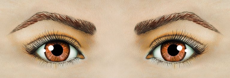
When we look at a person's face, the eyes we see are just a minute representation of the entire eyeball.
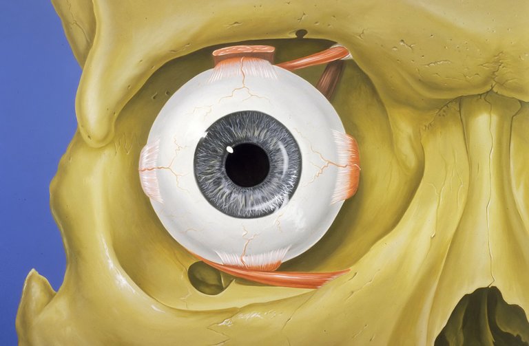
The eye is the organ of sight located in the orbital cavity. It is almost spherical in shape and its volume is approximately 7cc.

By Patrick J. Lynch, medical illustrator - Patrick J. Lynch, medical illustrator, CC BY 2.5, Wikimedia
Contained between the eyeball and orbital cavity is fatty yissue, which helps protect the eye from injury alongside the bony wall.
Though, structurally separate, the two eyes function as a pair.
It is possible to see with just one eye, but the three-dimensional vision is impaired when only one eye is used.

By Viktoria Borodinova - Own work, CC BY-SA 4.0, Wikimedia
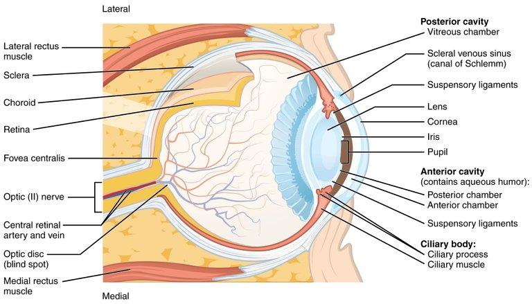
The eye is divided into layers and segments The layer encloses the cavities and is divided into;
- The outer fibrous layer, which comprises the Sclera, Cornea and Limbus.
- The middle vascular layer, comprises the Iris, Ciliary body, and Choroid.
- The inner nervous tissue layer, which comprises the Retina, Optic disc, and Optic nerve.
The segments includes;
- Anterior segment is further divided into Anterior and Posterior chamber.
The anterior segment contains the aqueous humor. - Posterior segment which contains the vitreous humor.

By OpenStax College - Anatomy & Physiology, Connexions Web site, Jun 19, 2013., CC BY 3.0, Wikimedia.
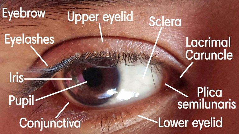
Having divided the eye into layers and segments, let's not forget the accessory structures of the eye, such as, eyebrow; eyelids and eyelashes; lacrimal apparatus; extraocular muscles of the eye.
Their main function being to protect the eye and help in movement of the eye.

By Biswadip Barman - Own work, CC BY-SA 4.0, Wikimedia
WHAT ABOUT THE EYE STRUCTURES?
Let us briefly describe these structures.
Sclera: a.k.a the white of the eye. It maintains the shape of the eye and gives attachment to the extraocular muscles. It is generally thick, except at the point where optic nerve pierces, called lamina cribrosa.
Cornea: forms the anterior 1/6 of the eye. This is the transparent anterior part of the eyeball and the main refracting surface of the eye, +43 to +45D.
Limbus: is the junction between the cornea and Sclera. A minute row of blood vessels about 1mm broad is present here.
Iris: is the part that gives various individual their eye colour (blue, brown, black e.t.c). It has an opening in the centre- the pupil.
Ciliary body: is triangular in shape with bade forwards. The iris is attached to the middle of the base. It consists of two main parts, pars plicata and pars plana.
Choroid: it lies between the Sclera and retina. It is dark brown and highly vascular, and extends to the optic nerve from the ora serrata.
Retina: lines about 3/4 of the eyeball. At the posterior part of the retina is a yellow area, called macula lutea, with a central depression, called fovea centralis (the most sensitive part of the retina).
Optic disc: has only nerve fibre later , so it doesn't excite any visual response. It is known as the blind spot.
Aqueous humor: is a clear fluid contained in the anterior segment. It is secreted into the posterior chamber by the ciliary epithelium, and moves into the anterior chamber via the pupil.
Lens: is transparent, circular, biconvex structure lying just behind the pupil. It is suspended from the ciliary body by suspensory ligament.
Vitreous humor: is also colorless transparent gel which fills the posterior segment (which is 4/5 of the eyeball). It consists of 99% water, some salts and mucoproteins.
BLOOD AND NERVE SUPPLY TO THE EYE
Internal carotid artery gives off ophthalmic artery, which gives off Ciliary (long and short) and central retinal arteries.
The Ciliary arteries and central retinal artery supply the eye.
Venous drainage is done by short Ciliary vein, anterior Ciliary veins, 4 vortex veins, and the central retinal vein. All these finally drain into the cavernous sinus.
The eye is supplied by 3types of nerves, namely motor, sensory and autonomic.
The motor nerves include, oculomotor, trochlear, abducens and facial nerve.
The trigeminal nerve is responsible for sensory supply.
The autonomic nerves are further divided into sympathetic (through the cervical sympathetic fibres), and parasympathetic (through the nuclei in the midbrain).
The sympathetic and parasympathetic fibres supply the iris (dilator pupillage muscle- sympathetic, sphincter pupillage muscle- parasympathetic), Ciliary body, and lacrimal glands.
REFERENCE
Congratulations @beulah4real! You have completed the following achievement on the Hive blockchain and have been rewarded with new badge(s):
Your next target is to reach 700 upvotes.
You can view your badges on your board and compare yourself to others in the Ranking
If you no longer want to receive notifications, reply to this comment with the word
STOPCheck out the last post from @hivebuzz: